


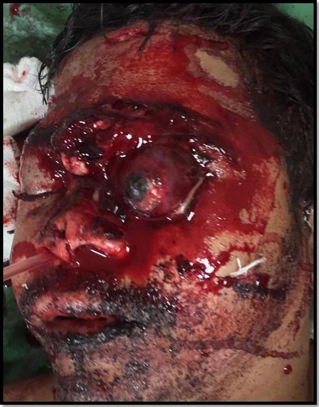
Image 1: Left sided eyeball subluxation and extensive craniofacial injury.

Image 2: Stepwise evacuation of retro-orbital hematoma. (a) Muscle identification and tagging. (b) Lateral canthotomy and inferior cantholysis. (c) Palpating optic nerve and orbital contents. (d) Orbital hematoma removal.

Image 3: Steps for globe reposition. (a) Ocular surface covered with amniotic membrane graft. Upper lid blepharotomy to lengthen anterior lamella and facilitate lid closure. (c) Placing Symblepharon ring. (d) Using symblepharon ring to mobilize globe and slip it under the upper lid.

Image 4: Surgical repair of craniofacial trauma. (a) Fixing palatal split. (b) Frontal fracture with CSF. (c) Harvesting Tensor fascia lata for dural repair. (d) On table outcome after tarsorrhaphy and wound closure.

Image 5: (a) Post operative appearance after 2 months. (b) Left eye prephthisical. Wound dehiscence of lower lid laceration repair.


 Slavshit

 Sandshit

Jump in the discussion.
No email address required.
Jump in the discussion.
No email address required.
More options
Context