


- 11
- 7
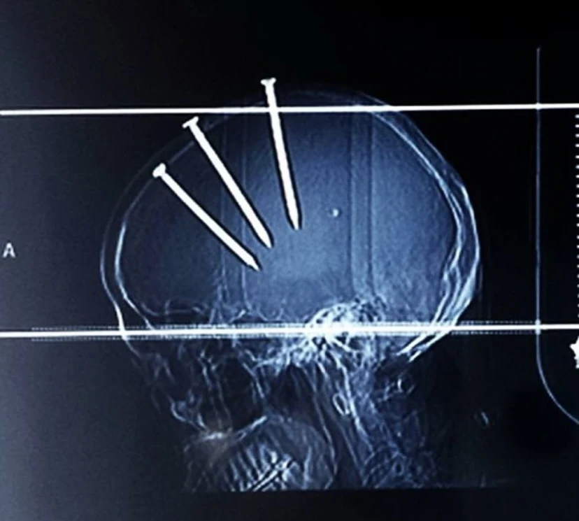
I think he had broken his toes or sum if anyone may know, correct me if i'm wrong 

- 4
- 26
If this is a repost imma kill myself


- 44
- 129
The Radium Girls were a group of female factory workers who tragically contracted radiation poisoning while working with radium-based paint. Here's a short and easy to digest outline of their story:
1. Background
Between 1917 and the early 1920s, these women worked at three different factories in the United States: one in Orange, New Jersey, another in Ottawa, Illinois, and a third in Waterbury, Connecticut. Their job involved painting watch dials and hands with a self-luminous paint containing radium. This paint allowed the watch faces to shine very bright light in the dark.
2. Innocent Beginnings
The women were initially told that the paint was harmless. To create fine brush tips, they were instructed to point their brushes on their lips, inadvertently ingesting deadly amounts of radium. Some were even allowed to paint their fingernails, faces, and teeth with the glowing substance after a long day of work.
3. Radium Poisoning
The paint was made from powdered radium, zinc sulfide, gum, and water. Despite being aware of the injurious effects of radium, the factory owners misled the workers. The women suffered from radiation exposure, leading to various diseases such as cancer, ulcers, and bone fractures. Many lost teeth, and experienced shattered bones. Their skin glowed for weeks and months after ingesting radium.
4. Legal Battles and Impact
In New Jersey, five women challenged their employer in court, seeking the right to sue for occupational diseases. They settled out of court in 1928. In Illinois, five women employed by the Radium Dial Company (unaffiliated with the United States Radium Corporation) won damages in 1938. The case of the "Radium Girls" significantly influenced workplace safety laws in the United States.
These brave women fought for their rights and safety, leaving a lasting impact on labor laws and raising awareness about the dangers of radium exposure. Their tragic story serves as a reminder of the importance of protecting workers' health and well-being.
Sources:
(1) Radium Girls - Wikipedia. https://en.wikipedia.org/wiki/Radium_Girls.
(2) Celebrating the Radium Girls' Fight Against Forever Chemicals This .... https://www.sierraclub.org/articles/2021/03/radium-girls-womens-history-month.
(3) How the 'Radium Girls' Helped Shape American Labor Laws - HistoryNet. https://www.historynet.com/radium-girls-vs-us-radium/.
(4) Radium Girls: The Women Who Fought for Their Lives in a Killer .... https://www.britannica.com/story/radium-girls-the-women-who-fought-for-their-lives-in-a-killer-workplace.
I highly suggest "Episode 176: Torture Deaths of the 'Ghost Girls'," a deep-dive from the Rotten Mango podcast. The link can be found below.


edit: omg thank you for all of the upvotes!
another edit: TYSM @Less-


")
- 2
- 11
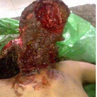
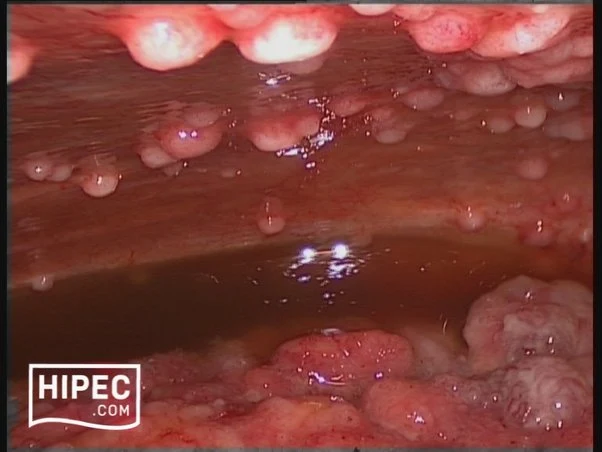
Here's an interesting case of subdural hematoma. In simpler terms, it's when blood leaks from your vessels into the space between the brain and the skull. Quite a spectacle, isn't it? As you can imagine, nothing good comes out of it.


- 46
- 115
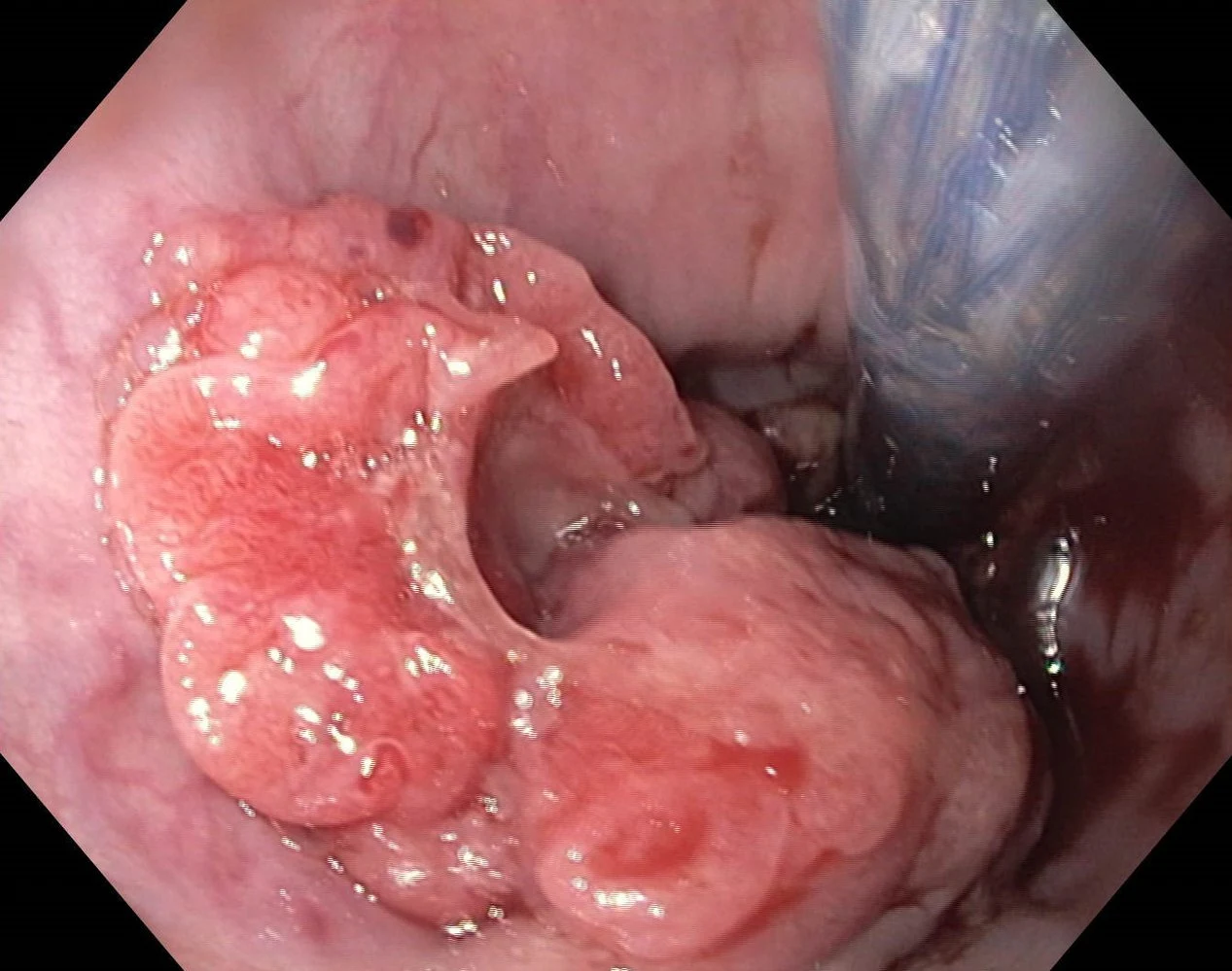
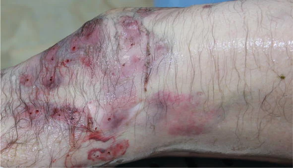
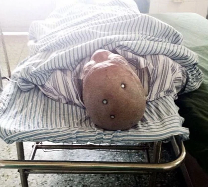
Fibrodysplasia ossificans progressiva is a connective tissue disorder, in which fibrous connective tissue, like ligaments, tendon and muscle are replaced by bone, causing the patient to stiffen up. This is the only medical condition known to cause one organ to grow into another.


Fibrodysplasia ossificans progressiva, or FOP, is extremely rare and estimated to affect 0.5 cases per million people. In 2017 there were only 801 cases known worldwide.
The cause of this disorder is a mutation of the gene, ACVR1. It is the body's repair mechanism, making it use bone to repair instead of the normal tissue. This causes joins to fuse and become immovable if even minor trauma occurs.
The new bone formation is called "heterotopic ossification," that new skeleton eventually restricts the patients ability to move, keeping them in a fixed position for the rest of their life.


Even if the excess bone is surgically removed, the body will repair itself with what is refered to as an explosive growth of new bone. There is no cure for this condition.
__________________
Hi this is my first post 
I've always been interest in medicine and crazy medical conditions so I'll probably make more! Don't know if this is an effortpost but that would be so cool if it was 


- 102
- 330


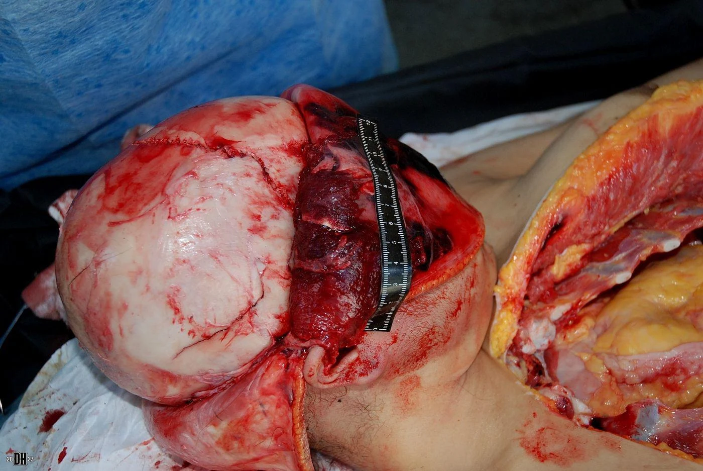

In the first video, there is hemorrhaging of the brain. Those black clots are the blood and they are dark because they began drying once the body died. When bodies are dead for a certain amount of time before the autopsy, many parts turn black, blue, or purple. Areas with more blood turn black, while more boney areas turn blue.
I personally think the human body is very beautiful with all the colors it can reveal after death. Every color of the rainbow can be found in the body in some way, although green is less common.


- 9
- 31
First of all who is Dr tsokos?

Michael Tsokos (born January 23, 1967 in Kiel) is a German forensic doctor and professor at the Charité in Berlin. He headed the Institute for Forensic Medicine at the Charité from 2007 to 2023 and at the same time he has headed the State Institute for Forensic and Social Medicine in Berlin-Moabit since 2007.
(you can find his instagram under dr.Toskos)

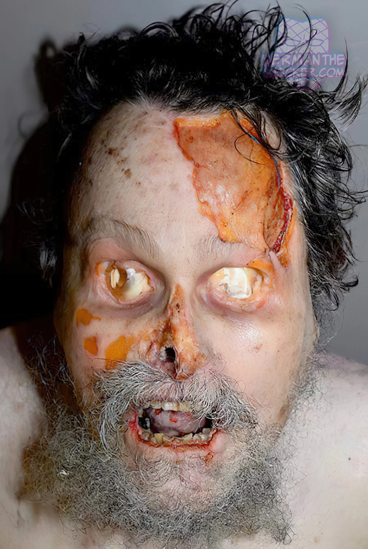
Here you can see an decapitated head preserved. The Victim committed suicide in the early to mid 19 hundreds.


Victim suffered a pulmonary embolism
Overview: A pulmonary embolism is a blood clot that blocks and stops blood flow to an artery in the lung. In most cases, the blood clot starts in a deep vein in the leg and travels to the lung. Rarely, the clot forms in a vein in another part of the body.


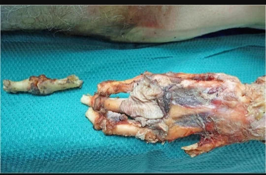
Hand and organs of a Victim that died due hypothermia
Overview: Hypothermia is a condition that occurs when core body temperature drops below 95 degrees Fahrenheit (35 degrees Celsius). It is a medical emergency. In hypothermia, the body loses heat faster than it can produce heat, causing a dangerously low body temperature.

Close up of a brain
Cause of death unknown

Heavy decomposed Body that was found in a moist basement
Cause of death unknown


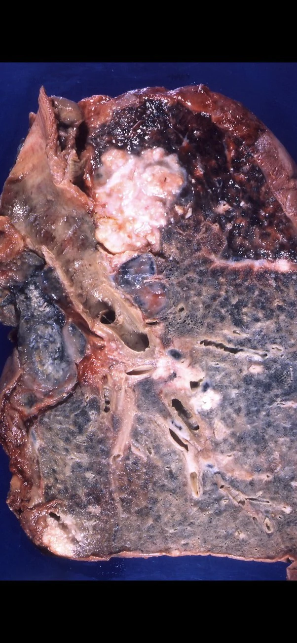
cirrhosis of the liver
Overview: Cirrhosis of the liver is a type of liver damage where healthy cells are replaced by scar tissue. The liver is unable to perform its vital functions of metabolism, production of proteins, including blood clotting factors, and filtering of drugs and toxins.

Preserved hand of a potential homicide victim with defends wounds (cuts).

Discoloration of the lymph nodes due to tattoo ink
Cause of death unknown

Skeletal remains. Victim dead for about 9 months (last time they‘ve been seen)
Cause of death unknown

X-ray of a dismembered body
Cause of death homocide
That's it for now 


- 5
- 27


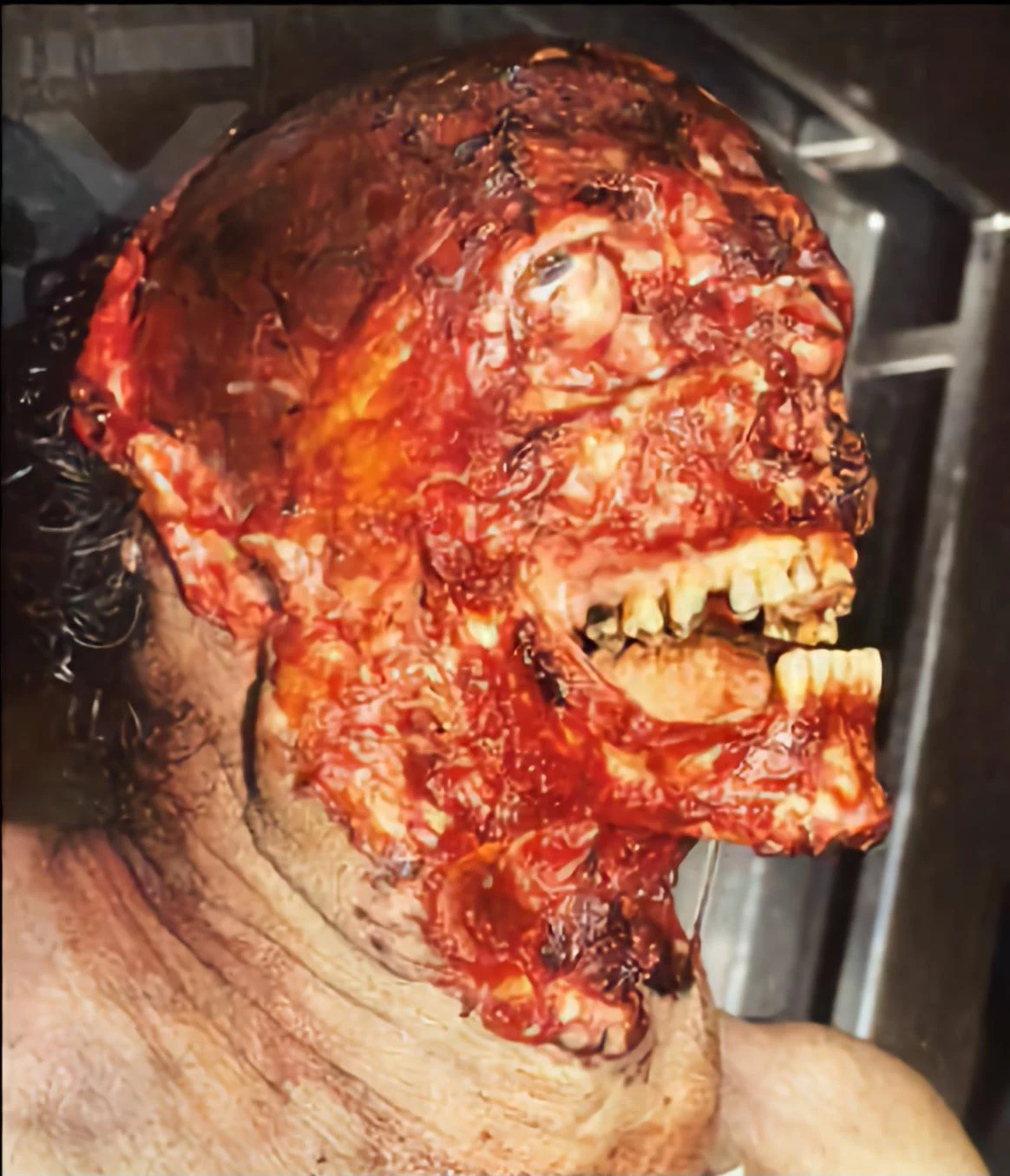
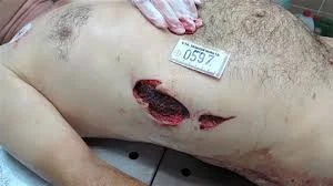
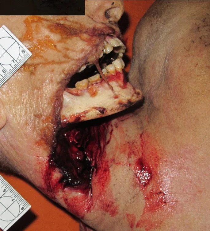
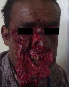
A 54-year-old male arrived at the regional trauma center following a self-inflicted gunshot wound to the face (Figure 1).
The defect spanned the left side of the face inferolateral to the nose and inferior to the left eye. The defect had abraded and jagged edges. After stabilization, tracheostomy placement, and two rounds of debridement, a vascularized free fibula reconstruction was planned and performed using 3D modeling (Figure 2).
The intraoral defect was reconstructed using local tissues but dehisced following multiple attempts at intraoral closure. The initial closure attempts consisted of (1) local tissues at the time of initial flaps from the floor of the mouth and buccal lining tissue, followed by (2) spanning the defect with a thin piece of acellular dermal matrix (ADM) at a second operation, and (3) subsequently with another piece of ADM covered with a pedicled floor of mouth flap. These local flaps failed, exposing fibular bone and hardware. At the fourth operation, a split skin paddle STAIF was used for extra- and intra-oral coverage that led to stable coverage.
The design of the STAIF begins with identifying the superficial temporal artery (STA) via a Doppler probe. Typically, the STA is easily palpated approximately 2 cm anterior to the anterior helical rim and courses superiorly. In our experience, the flap may be elevated at least 5 cm past the last Doppler-able vessel location. After identifying the vascular course, the skin is pinched to visualize how much can be closed primarily. Marks are made to accommodate the primary closure of the donor site.
The skin paddle is incised and elevated in a superior to inferior direction. Additional temporoparietal (TP) fascia, galea, or pericranium may be included via undermining for more extensive coverage or fascial wrapping of the fibula or hardware. In this case, the TP, STA, and fascia were dissected down to the level of the temporalis muscle, keeping the pedicle with the flap. Dissection was maintained posterior to the course of the frontotemporal branch of the facial nerve, which lies immediately deep to the TP fascia. The skin paddle was islandized, and the flap elevated to the level of the zygomatic arch. A tunnel was made in the cheek superficial to the zygomatic arch at the level of the tragus. The flap was passed into the mouth, crossed the midline of the mandibular gingivobuccal sulcus, and sutured intraorally to cover the gingivobuccal sulcus of the lower lip and mucosal defect in the floor of the mouth (Figure 3).
For more extended reach, the flap may be passed under the zygomatic arch. In this case, the skin paddle was divided and split, providing both intra- and extra-oral coverage, like the methods described by Elbanoby et al. [3]. The distal skin paddle served for intraoral defect coverage; the proximal allowed for the reconstruction of the cheek and outer mouth after partial de-epithelization. The external facial defect was in the beard line and reconstructed with hair-bearing skin. The donor site was closed primarily after wide undermining and healed favorably. A Doppler signal was identifiable in each skin paddle.
Intraoral competence was restored following the STAIF use in this patient. Coverage of the fibula and hardware was obtained without further dehiscence or exposure (Figure 4).
He returned two months postoperatively for debulking and flap inset given an element of microstomia and difficulty with lip elevation. The flap was further divided at its tip into two portions for improved intra- and extraoral delineation and enhanced lip commissure definition. The split skin paddle STAIF successfully reconstructed intraoral defects and external cheek soft tissue wounds (Figures 5, 6).
Top Poster of the Day:

 FewerBeary
Deaths Today: 0
Current Registered Users: 2,091,395
 BROWSE EFFORTPOSTS
SITE GUIDE
PING GROUPS
BROWSE EFFORTPOSTS
SITE GUIDE
PING GROUPS
Medical

Aspiring medical student? Or maybe just have an interest in human anatomy? Here you will find everything from autopsies to surgeries to photos from medical journals. If it happened in the operating room or the morgue, it probably belongs here.
 Slavshit
 Sandshit