


 Posting medical gore is my thing
- 2
- 14
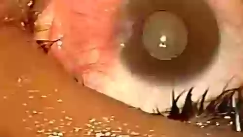
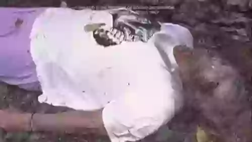
An eye web is a noncancerous, triangular growth that may occur on one or both eyes. It's more common in people who spend a lot of time in the sun, such as those who work outdoors.
The painless growth may be slightly raised and contain obvious blood vessels. It may cause irritation and possibly affect vision.
Treatment usually isn't necessary. Eyedrops or surgery may help in severe cases.


- 3
- 27
from the description:
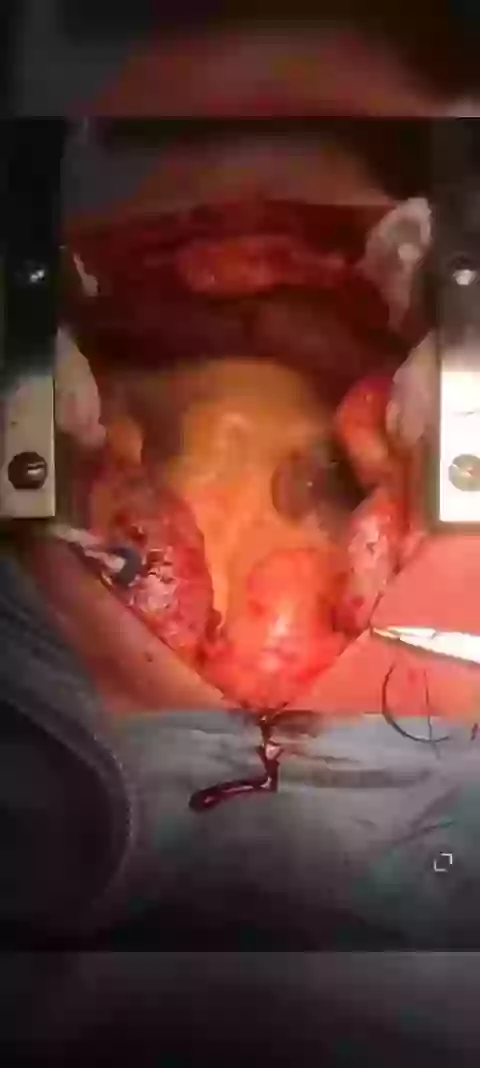
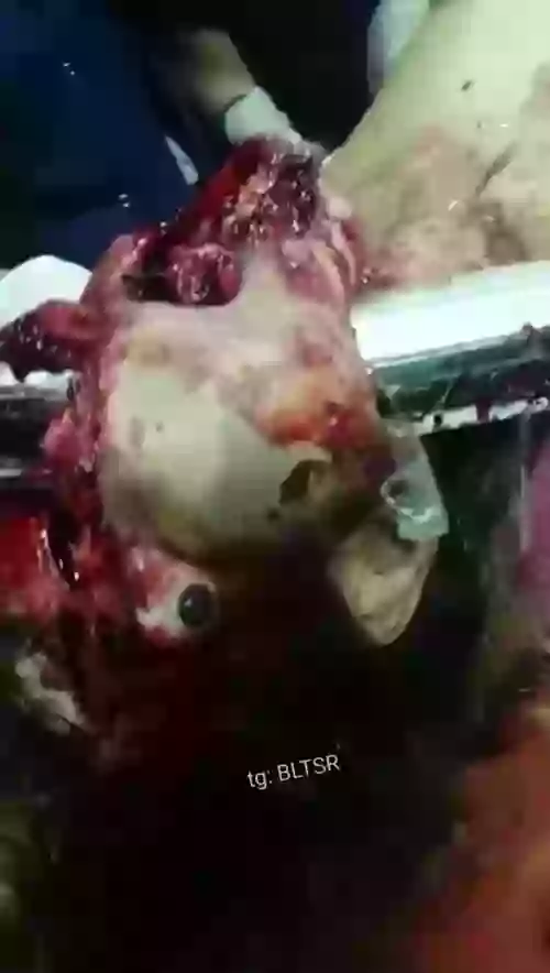
Video describes how a total thyroid removal is performed along with possible complications. Specifically discussed are recurrent laryngeal nerve damage causing permanent hoarseness as well as parathyroid gland damage leading to calcium regulation problems in the body.
For more information on thyroid surgery: https://www.fauquierent.net/thyroidmass.htm
Video produced by Dr. Chris Chang: https://www.FauquierENT.net
- 19
- 67
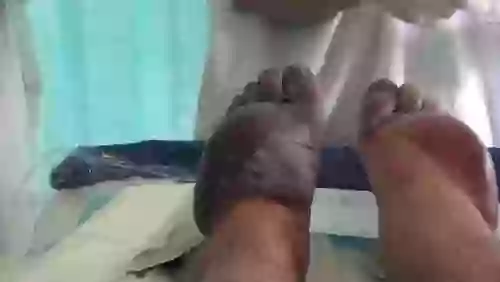
Skin jiggers, also known as chigoe fleas or sand fleas, are small parasitic insects that burrow into the skin, typically on the feet and legs. They are common in tropical and subtropical regions and can cause severe itching, inflammation, and infection. They can be treated with topical medications, but preventing infection by avoiding contact with the fleas is the best approach.


- 2
- 21
from the description:
this 59-year old patient had a history of long standing exotropia with limited horizontal movements of the eye. it’s unclear if she had it from birth but probably had it from her early childhood. dr. wagner maximally recessed the lateral rectus muscle and resected the medial rectus muscle. he performed the surgery in both eyes but only one eye surgery is demonstrated here.
surgeon: dr. rudolph wagner, rutgers – new jersey medical school, newark, nj, usa

- 9
- 30
BACKGROUND:
Clubfoot occurs in approximately one in 1000 live births and is one of the most common congenital birth defects. Although there have been several reports of successful treatment of idiopathic clubfoot with the Ponseti method, the use of this method for the treatment of other forms of clubfoot has not been reported. The purpose of the present study was to evaluate the early results of the Ponseti method when used for the treatment of clubfoot associated with distal arthrogryposis.
METHODS:
Twelve consecutive infants (twenty-four feet) with clubfoot deformity associated with distal arthrogryposis were managed with the Ponseti method and were retrospectively reviewed at a minimum of two years. The severity of the foot deformity was classified according to the grading system of Diméglio et al. The number of casts required to achieve correction was compared with published data for the treatment of idiopathic clubfoot. Recurrent clubfoot deformities or complications during treatment were recorded.
RESULTS:
Twenty-two clubfeet in eleven patients were classified as Diméglio grade IV, and two clubfeet in one patient were classified as Diméglio grade II. Initial correction was achieved in all clubfeet with a mean of 6.9 +/- 2.1 casts (95% confidence interval, 5.6 to 8.3 casts), which was significantly greater than the mean of 4.5 +/- 1.2 casts (95% confidence interval, 4.3 to 4.7 casts) needed in a cohort of 219 idiopathic clubfeet that were treated during the same time period by the senior author with use of the Ponseti method (p = 0.002). Six feet in three patients had a relapse after initial successful treatment. All relapses were related to noncompliance with prescribed brace wear. Four relapsed clubfeet in two patients were successfully treated with repeat casting and/or tenotomy; the remaining two relapsed clubfeet in one patient were treated with extensive soft-tissue-release operations.
- 6
- 23
Eye.
A skin graft is a surgical procedure in which a piece of healthy skin is removed from one area of the body and used to repair or cover a damaged area of skin. There are several different types of skin grafts, each with its own advantages and disadvantages.
Split-thickness skin graft: This is the most common type of skin graft. It involves removing the top layers of skin (epidermis and part of the dermis) from a healthy area of the body, called the donor site, and transplanting it to the damaged area, called the recipient site. The donor site is usually closed with stitches or staples and will heal within a few weeks. The grafted skin will eventually blend in with the surrounding skin and provide a protective barrier for the damaged area.
Full-thickness skin graft: This type of skin graft involves removing the entire thickness of skin from the donor site and transplanting it to the recipient site. Because the entire thickness of skin is removed, the donor site requires a more extensive surgical procedure and a longer recovery time. The grafted skin will blend in with the surrounding skin, but will not have any hair follicles or sweat glands, which may affect its appearance and function.
Composite skin graft: This type of skin graft involves removing a combination of skin, fat, and muscle from the donor site and transplanting it to the recipient site. This type of graft is used to repair deeper burns or other injuries that have damaged the underlying tissue. Because the graft includes not only skin, but also fat and muscle, it will have a more natural appearance and better function.
Pedicle skin graft: This type of skin graft involves removing a piece of skin from the donor site and leaving it attached to the underlying tissue. The skin is then moved to the recipient site and sutured in place, allowing blood vessels to continue to nourish the grafted tissue. This type of graft is used when the recipient site is near the donor site and when the recipient site is too large to be covered by the skin available at the donor site.
Allograft: This type of skin graft uses skin from a donor of the same species (human) but different individual. The allograft can come from a cadaver, or from a living individual such as in a case of a burn victim. The allograft is cleaned and sterilized, and then applied to the recipient site.
Xenograft: This type of skin graft uses skin from a different species, such as pig or cow. The xenograft is cleaned and sterilized, and then applied to the recipient site. While xenografts can be used as a temporary covering for severe burns, the body can react to the foreign tissue, requiring additional treatment.
It's important to note that skin grafts can be associated with some risks, such as infection, bleeding, and rejection of the grafted tissue. Also, the success of the graft depends on the health and condition of the patient, the size and location of the wound, and the availability of healthy skin at the donor site. It's essential to have a good follow-up after the surgery and to take care of the grafted area to avoid complications.
- 3
- 45
Burns are injuries to the skin and underlying tissues caused by heat, chemicals, electricity, or radiation. The severity of a burn is classified based on its depth and extent, which determines the appropriate treatment. The stages of burn damage to human tissue are as follows:
First-degree burn: This is the mildest form of burn, and affects only the outer layer of skin (epidermis). Symptoms include redness, pain, and mild swelling. Sunburn is an example of a first-degree burn. Treatment includes cleaning the burn and covering it with a sterile bandage, over-the-counter pain relievers, and cool compresses to reduce pain and inflammation.
Second-degree burn: This burn affects the epidermis and the underlying layer of skin (dermis). Symptoms include redness, pain, swelling, and blisters. Treatment includes cleaning the burn, covering it with a sterile bandage, over-the-counter pain relievers, and cool compresses to reduce pain and inflammation. It's important to cover the burn to prevent infection. Second-degree burns usually heal within 2-3 weeks.
Third-degree burn: This burn affects the epidermis, dermis and underlying tissue (subcutaneous tissue). Symptoms include white or blackened, charred skin that may be numb to the touch. Treatment includes cleaning the burn, covering it with a sterile bandage, over-the-counter pain relievers, and cool compresses to reduce pain and inflammation. Third-degree burns may require skin grafts and plastic surgery to heal.
Fourth-degree burn: This is the most severe form of burn, affecting not only the skin and underlying tissue, but also the bones, muscles, and tendons. Symptoms include blackened, charred skin, severe pain, and possible damage to nerves and muscles. Treatment includes cleaning the burn, covering it with a sterile bandage, over-the-counter pain relievers, and cool compresses to reduce pain and inflammation. Fourth-degree burns may require skin grafts and plastic surgery, as well as amputation in some cases.
It's important to note that in case of severe burn, immediate medical attention is necessary. If a burn is larger than 3 inches or is on the face, hands, feet, or a major joint, it should be considered as a severe burn and professional medical help should be sought.
- 10
- 66
Frostbite is a condition caused by freezing of the skin and underlying tissues. It typically affects the extremities, such as the fingers, toes, nose, and ears. The stages of frostbite are as follows:
Frostnip: This is the earliest stage of frostbite. The skin appears white or blue and is numb, but can still be warmed back to normal color and temperature. This stage is considered mild and reversible, and can be treated with rewarming and protection from further cold exposure.
Superficial frostbite: In this stage, the skin becomes hard and pale and blisters may appear. The affected area is numb and there is little or no pain. The tissue is still soft and can be easily pinched. The damage in this stage is considered reversible with proper treatment.
Deep frostbite: In this stage, the skin becomes black or purple and appears waxy. The affected area is numb and the tissue is hard and frozen. Blisters may appear, and the tissue may be destroyed. The damage in this stage is considered irreversible and may require amputation.
Symptoms of frostbite include:
Numbness or tingling
Hard or waxy-looking skin
Blisters
Swelling
Discoloration (white, blue, black)
Loss of feeling in the affected area
Loss of movement in the affected area
Treatment for frostbite includes rewarming the affected area in warm water (104-108 degrees Fahrenheit) and protecting it from further cold exposure. Pain medication may be given for the discomfort. In severe cases, antibiotics may be prescribed to prevent or treat infection. Hyperbaric oxygen therapy (HBOT) may also be used to increase oxygen levels to the affected tissue, which can aid in healing. In some cases, amputation may be necessary if the tissue is severely damaged and cannot be saved.
It's important to note that the earlier the frostbite is treated, the better the chance of recovery. In addition, preventing frostbite by keeping the body warm and dry, wearing appropriate clothing and avoiding prolonged exposure to cold temperatures, are key to avoiding frostbite.
@Zin here's your frostbite video!
- 12
- 110
The decomposition of the human body can be divided into several stages. These stages are influenced by environmental factors such as temperature, humidity, and the presence of insects or other organisms.
Fresh stage: This is the initial stage of decomposition, which occurs immediately after death. The body is still relatively intact and appears normal, with a natural pallor due to the loss of blood circulation.
Early decomposition: This stage occurs within the first few days after death. The body begins to cool and becomes stiff, a process known as rigor mortis. As bacteria within the body begin to break down tissues, the body may develop a greenish or purplish discoloration known as livor mortis.
Bloat stage: This stage occurs within the first week after death. Bacteria within the body continue to break down tissues, producing gases that cause the abdomen to swell and the skin to take on a discolored, mottled appearance.
Active decay: This stage occurs within the first two to three weeks after death. The body begins to liquefy as bacteria continue to break down tissues, causing a strong, foul odor. Maggots and other insects may be present at this stage.
Advanced decay: This stage occurs within several weeks to several months after death. The body is now mostly bones and hair, and the odor is less intense.
Dry/skeletonization: This is the final stage of decomposition, which can take several months to several years, depending on environmental factors. The body is now reduced to dry bones and hair.
It's worth noting that decomposition can be slowed down by embalming process and it's not always follow the exact stages described above. Also, the stages of decomposition may be different for bodies that are buried underground and those that are exposed to the elements.


- 31
- 73


- 5
- 17
Segment 1 The procedure for simple extraction of cataracts is shown. The procedure is repeated on many eyes, and the films have been edited together in a series. The puncture of the eyeball, incision of capsule and counterpuncture are shown in the procedure. An example of a difficult extraction and an example of an easy extraction are shown. The procedure for extracting a secondary cataract with a needle is shown. The method of extraction by a buccal vacuum and suction is shown.
Segment 2 A series of cataract extractions continues to be shown. Then the method for irrigating the anterior chamber of the eye and replacing the iris is shown, again in a series of operations

- 7
- 52
Necrosis is a pathological process characterized by the death of cells or tissues in the body. It can occur as a result of various etiologies, including ischemia, trauma, infection, inflammation, and exposure to toxins, among others. The various types of necrosis can be classified based on the morphological changes that occur in the affected cells and tissues, as well as the underlying mechanisms of cell death.
Coagulative necrosis is one of the most common types of necrosis and is characterized by the coagulation of proteins within the affected cells and tissues. This type of necrosis typically occurs as a result of ischemia, in which blood flow to a specific tissue or organ is abruptly cut off. The affected tissue becomes firm and hard, and appears white or yellow in color, a phenomenon known as "coagulative infarction". This type of necrosis is commonly observed in myocardial infarction and cerebral infarction. Treatment for coagulative necrosis centers on preventing further damage and supporting organ function. After a heart attack, healthcare providers initially focus on restoring blood flow to your heart, if possible. They also work to reestablish normal heart function.

Liquefactive necrosis is another type of necrosis, characterized by the liquefaction of the affected cells and tissues. This occurs as a result of the hydrolytic enzymes released by neutrophils and macrophages, which break down the cellular components and lead to the formation of a liquid or pus-like substance. This type of necrosis is commonly observed in abscesses, pyogenic infections, and certain types of brain tumors.Treatment of liquefactive necrosis typically involves two distinct processes. Usually, the underlying cause of the necrosis must be treated before the dead tissue itself can be dealt with. Even after the initial cause of the necrosis has been halted, the necrotic tissue will remain in the body. The body’s immune response to apoptosis, the automatic breaking down and recycling of the cell material, is not triggered by necrotic cell death. Antibiotics will be prescribed. Antibiotics that work against a number of different bacteria (broad spectrum antibiotics) are most commonly used. You may be prescribed several different types of antibiotics to make sure treatment works. Antifungal medications may also be prescribed if the infection is likely caused by a fungus. The standard therapy of liquefactive necrosis is surgical removal of necrotic tissue. Depending on the severity of the necrosis, this may range from removal of small patches of skin, to complete amputation of affected limbs or organs. Chemical removal, via an enzymatic debriding agent, is another option. In select cases, special maggot therapy has been utilized with good results.


Caseous necrosis is a type of necrosis characterized by the formation of a cheese-like substance within the affected cells and tissues. This type of necrosis is typically observed in granulomatous diseases, such as tuberculosis and sarcoidosis. In this type of necrosis, the cells die and form a caseous material, surrounded by a layer of inflammatory cells known as "epithelioid cells" and "Langhans giant cells". Healthcare providers treat the conditions that cause caseous necrosis. TB treatment is usually several months long and may involve multiple medications. This is because TB can be antibiotic resistant. Your provider will establish a treatment regimen for you and help you manage side effects from the medications.


Fibrinoid necrosis is a specific form of necrosis that occurs in blood vessels. This type of necrosis is characterized by the deposition of fibrin and other proteins within the walls of the vessels, leading to thickening and narrowing of the lumen. This type of necrosis is commonly observed in autoimmune diseases, such as lupus and rheumatoid arthritis. Can result in bruising and darkening of the infected area. In general, there’s no standard treatment. But healthcare providers can treat bleeding. This depends on the type of bleeding and where it is in the body. Providers can also treat the conditions that cause fibrinoid necrosis. For example, medications can lower blood pressure or suppress the immune system and limit further damage to the blood vessels.


Fat necrosis is a type of necrosis that occurs in adipose tissue, characterized by the breakdown of fat cells. This type of necrosis can occur as a result of injury or inflammation and is commonly observed in the breast tissue, pancreas, and skin. Most people do not need treatment. Fat necrosis is harmless so you will not usually need any treatment or follow-up. In most cases the body will break it down over time. This could take a few months. However It’s important to go back to your GP if the lump gets bigger or you notice any other changes to your breasts.

(breat tissue affected)
Gangrenous necrosis is a type of necrosis characterized by the death of tissue in a specific area, often as a result of a lack of blood supply. This type of necrosis is commonly observed in severe infections, such as sepsis, and in cases of peripheral artery disease. Treatments for gangrene may include antibiotics, oxygen therapy, and surgery to restore blood flow and remove dead tissue. The earlier gangrene is identified and treated, the better the chances for recovery.


In conclusion, necrosis is a pathological process that can occur in any tissue or organ and can result in permanent damage or loss of function. Understanding the different types of necrosis, their causes, and morphological changes can aid in the diagnosis and treatment of various diseases and injuries. It is important to seek medical attention if necrosis is suspected, as it can be a sign of a serious underlying condition.


- 36
- 37
Nurafaiha was born with a cleft lip and pallet as well has having no skull and is blind. Her disabilities mean that she cannot take milk normally and has to be fed through a tube.
https://www.mirror.co.uk/news/world-news/baby-born-without-skull-half-9511769
- 18
- 100
A 8 year old girl presented with a history of Pencil Injury. She has a broken pencil lead stuck inside her cornea traversing into the anterior chamber.

Top Poster of the Day:
 Boydicker
Deaths Today: 0
Current Registered Users: 2,027,672
 BROWSE EFFORTPOSTS
SITE GUIDE
PING GROUPS
BROWSE EFFORTPOSTS
SITE GUIDE
PING GROUPS
Medical

Aspiring medical student? Or maybe just have an interest in human anatomy? Here you will find everything from autopsies to surgeries to photos from medical journals. If it happened in the operating room or the morgue, it probably belongs here.
 Slavshit

 Sandshit